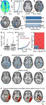
Chronic traumatic encephalopathy (CTE), a neurodegenerative disease caused by repeated head injuries often affecting athletes, can only be diagnosed currently through brain tissue analysis post-mortem. However, in a new study published in Brain, a Journal of Neurology, Ben-Gurion University of the Negev (BGU) researchers present a new test methodology. Using brain imaging techniques and analytical methods, they can determine whether football players have CTE by measuring leakage of the blood-brain barrier.
The blood-brain barrier (BBB) is a specialized interface between the blood and the brain environment that prevents the transfer of unwanted molecules or infectious organisms from the blood to the brain. Evidence shows that breaching the integrity of this barrier causes many brain diseases and neurodegeneration as a result of aging.
“Since a leaky BBB is also found in CTE and causes brain dysfunction and degeneration, it now seems that this test could provide the first (and so far the only) evidence for brain injury in the players we studied on the Israel football team,” says Prof. Alon Friedman, M.D., a neurosurgeon and researcher at BGU and Dalhousie University in Canada.
“Importantly, we believe that those with persistent leak encompassing months or years are more likely to develop CTE. Many players seem to repair their BBB quickly, and if they do not suffer from repeated TBIs [traumatic brain injuries] or are not sensitive to brain injury, they are not likely to develop CTE.”
The study population included 42 Israelis who play amateur American football in the Israeli Football League (IFL), and a control group comprising 27 athletes practicing a non-contact sport and 26 non-athletes. Magnetic resonance scans were also performed on 51 patients with malignant brain tumors, ischemic stroke or traumatic brain injury (TBI). The NFL sideline concussion assessment tool was used to document history of previous head injuries, including concussions, as well as symptoms assessment and Standardized Assessment of Concussion (SAC) tests.
The researchers developed a modified dynamic contrast-enhanced-MRI (DCE-MRI) protocol and analytical methods to investigate vascular pathology and blood-brain barrier disorder (BBBD) associated with repeated mild TBI in American football players. For the first time, using human brain imaging, they distinguished between fast and slow leakage through the pathological BBB and showed that localized, specific post-traumatic vascular pathology may persist for months in a subset of players.
“We generated maps that visualized the permeability value for each 3-D section (voxel), Prof. Friedman says. “Our permeability maps revealed an increase in slow blood-to-brain transport in a subset of amateur American football players, but not in the control group. The increase in permeability was region specific (white matter, midbrain peduncles, red nucleus, temporal cortex) and correlated with alterations in white matter tracts. Importantly, increased permeability persisted for months, as seen in players who were scanned both on- and off-season.
“Not less important is the observation that few players who did not complain of severe symptoms also showed a leaky BBB. This suggests that DCE-MRI should be used in conjunction with symptom questionnaires before return to play is approved.”
Football players were three times more likely to display a leaky BBB than controls as BBBD was detected in a subgroup (27.4%) of players. This individual variability may explain the wide range of cognitive deficits and neuropsychiatric impairments observed in players, likely reflecting differences in affected brain networks.
“Our findings show that DCE-MRI can be used to diagnose specific vascular pathology after traumatic brain injury and other brain pathologies,” Prof. Friedman says.
Source: Read Full Article
